


Blocked cell wall formation stops bacterial cell division

Blocked cell wall formation stops bacterial cell division according to researchers from University Hospital Bonn, Germany
The researchers say they have clarified the inhibitory effect mechanism of antibiotics on the growth of the bacterium, Staphylococcus aureus.
We still do not understand exactly how antibiotics kill bacteria. However, this understanding is necessary if we want to develop new antibiotics. And that is precisely what is urgently needed, because bacteria are currently showing more and more resistance to existing antibiotics.
Therefore, researchers from the University Hospital Bonn (UKB) and the University of Bonn used high-performance microscopes to observe the effect of different antibiotics on the cell division ofStaphylococcus aureus.
They found that the biosynthesis of peptidoglycan, a core component of the bacterial cell wall, is the driving force during the entire process of cell division. In addition, they clarified how exactly different antibiotics block cell division within a few minutes.
The bacterial cell wall maintains the shape and integrity of unicellular organisms. Cell wall synthesis plays a key role in bacterial growth: the cell division protein FtsZ forms the so-called Z-ring in the centre of the cell, thus initiating the division process.
A new cell wall is formed there, for which peptidoglycan is produced as the core component. This constriction thus gives rise to two identical daughter cells.
See also: New project hope for next generation of antibiotics
Cell division protein
The UKB research team led by Fabian Grein and Tanja Schneider, together with the team led by Ulrich Kubitscheck, Professor of Biophysical Chemistry at the University of Bonn, selected the bacterium Staphylococcus aureus, one of the most dangerous human pathogenic bacteria, as the model organism for their study.
The focus was on the influence of antibiotics that inhibit peptidoglycan synthesis on cell division.
Jan-Samuel Puls, a PhD student at the Institute of Pharmaceutical Microbiology at UKB, said: “We found a rapid and strong effect of oxacillin and the glycopeptide antibiotics vancomycin and telavacin on cell division. The cell division protein FtsZ served as a marker here and we monitored it.”
For this purpose, FtsZ was fluorescently labelled alongside other proteins. Then the researchers analysed the effects on individual living bacterial cells over time and also used super-resolution microscopy.
They established an automated image analysis for microscopy images that allowed them to quickly analyse all cells in the sample under study.
Dr Fabian Grein, junior research group leader at the UKB’s Institute of Pharmaceutical Microbiology and a scientist at the German Centre for Infection Research (DZIF), said: “Staphylococcus aureus is only about one micrometre, which is one-thousandth of a millimetre. This makes microscopy particularly challenging.”
Crucial
The Bonn research team found that the formation of peptidoglycan is the driving force during the entire process of cell division. Previously, peptidoglycan synthesis was thought to be essential only during a specific part of this process.
The team showed that inhibition of cell wall assembly by glycopeptide antibiotics in Staphylococcus aureus occurs rapidly and with a dramatic effect on cell division.
In addition, they clarified in detail the specific role of essential penicillin-binding protein 2 (PBP2), which links cell wall components, in cell division. The β-lactam antibiotic oxacillin prevents the proper localisation of this protein.
Grein said: “This means that PBP2 does not get to the place where it is needed. As a result, the cell can’t divide.
“Importantly, this all happens immediately after the antibiotics are added, so the first cellular effects, which have not been studied very intensively so far, are crucial.”
Therefore, in view of the alarming increase in antibiotic resistance worldwide, he hopes the study results will provide a better understanding of how exactly these agents work at the cellular level, and thus a key to the development of new antibiotics.
Understanding cellular mechanisms of antibiotic action and production is the goal of the DFG Collaborative Research Centre TRR 261 ‘Antibiotic CellMAP’, which conducted these studies.
The results have now been published in the journalScience Advances.
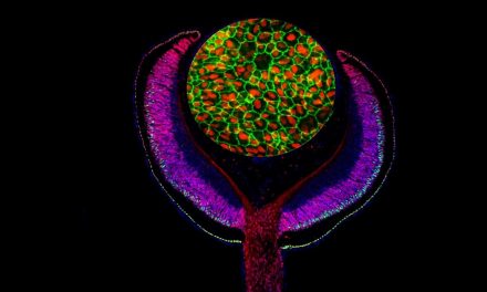
Image: Red fluorescent cell division protein FtsZ assembles so-called Z-ring in the centre of the cell. © University of Bonn/ Dominik Brajtenbach.


